Home
/ Diagram Of Liver Cell / Hepcludex First Drug For Hepatitis D Has Been Approved German Center For Infection Research : Form specific compounds such as coagulation factors and somatomedins or growth factors.
Diagram Of Liver Cell / Hepcludex First Drug For Hepatitis D Has Been Approved German Center For Infection Research : Form specific compounds such as coagulation factors and somatomedins or growth factors.
Diagram Of Liver Cell / Hepcludex First Drug For Hepatitis D Has Been Approved German Center For Infection Research : Form specific compounds such as coagulation factors and somatomedins or growth factors.. Portal tracts are channels that originate at the hilum and course through the liver in a branching pattern. The typical volume of a hepatocyte is 3.4 x 10 −9 cm 3. Parenchymal cells account for around 60% of the liver's structure. Liver cells are readily permeable to glucose. In humans, it is located in the right upper quadrant of the abdomen, below the diaphragm.its other roles in metabolism include the regulation of glycogen storage, decomposition of red blood cells, and the production of hormones.
The countless bile canaliculi join together into many larger bile ducts found throughout the liver. Smartdraw includes 1000s of professional healthcare and anatomy chart templates that you can modify and make your own. Functions of liver cells ƽ intricately involved in carbohydrate, fat, and protein metabolism. These substances are necessary for cell membrane production, digestion, bile acid formation, and hormone production. Draw a diagram of a liver cell ib biology syllabus ib biology 231 drawing a liver cell youtube ib biology notes 23 eukaryotic cells ib biology topic 231 drawing a liver cell youtube schematic representation of copper metabolism within a liver cell.
A Human Liver Cell Atlas Revealing Cell Type Heterogeneity And Adult Liver Progenitors By Single Cell Rna Sequencing Biorxiv from www.biorxiv.org It is a hugely important gland that is responsible for a wide range of metabolic and chemical reactions that are vital to living. Cell structure liver cell diagram wiring diagram database liver cell as an example of an animal cell diagram quizlet 231 draw and label a diagram of the ultrastructure of a liver cell as an example of an animal cell cell diagram 32 the cytoplasm and cellular organelles anatomy and physiology rough endoplasmic reticulum. Kupffer cells are irregular, with cytoplasmic extensions that facilitate their phagocytic function (fig. Human liver diagram with labels for exam point of perspective. Follow along and practise drawing a liver cell. Hemoglobin is degraded to iron and biliverdin, which is converted to bilirubin. The liver is thought to be responsible for up to 500 separate functions, usually in combination with other systems and organs. This helps carry away waste products from the liver.
The hepatic lobules are composed predominantly of liver cell trabecular cords one cell thick.
In humans, it is located in the right upper quadrant of the abdomen, below the diaphragm.its other roles in metabolism include the regulation of glycogen storage, decomposition of red blood cells, and the production of hormones. Bile produced by liver cells drains into microscopic canals known as bile canaliculi. In addition, there is a notable relationship between the number of afm papers and the frequency of occurrence of the different liver sinusoidal cell. The sinusoids are lined by phagocytic kupffer cells and hepatic stellate cells (or ito cells) inhabit the space of disse and function as. Parenchymal cells account for around 60% of the liver's structure. It's a huge organ that sits roughly in the middle of your abdomen. The countless bile canaliculi join together into many larger bile ducts found throughout the liver. Follow along and practise drawing a liver cell. The liver is thought to be responsible for up to 500 separate functions, usually in combination with other systems and organs. Excessive alcohol consumption can cause liver disease. Hepatocytes display an eosinophilic cytoplasm, reflecting numerous. Smooth endoplasmic reticulum is abundant in hepatocytes, whereas most cells in the body have only small amounts. Start studying ib biology topic 1.2:
The structure and function of liver cells. Smooth endoplasmic reticulum is abundant in hepatocytes, whereas most cells in the body have only small amounts. Uruj zehra mbbs, mphil, phd last reviewed: Hepatocytes display an eosinophilic cytoplasm, reflecting numerous. Hemoglobin is degraded to iron and biliverdin, which is converted to bilirubin.
Diagram Showing Different Stem Cell Applications Vector Image from cdn5.vectorstock.com The countless bile canaliculi join together into many larger bile ducts found throughout the liver. The liver is thought to be responsible for up to 500 separate functions, usually in combination with other systems and organs. In addition, there is a notable relationship between the number of afm papers and the frequency of occurrence of the different liver sinusoidal cell. Parenchymal cells account for around 60% of the liver's structure. Adrian rad bsc (hons) • reviewer: In this video i'm going to draw diagram of liver, stomach and pancreas labelled diagram from chapter human nutrition of class 11 biology.how to draw liver. Although an agent tends to cause initial damage in only one of these areas, the resulting disease may in time also involve other components. The hepatic lobules are composed predominantly of liver cell trabecular cords one cell thick.
These substances are necessary for cell membrane production, digestion, bile acid formation, and hormone production.
The typical volume of a hepatocyte is 3.4 x 10 −9 cm 3. The tubes that carry bile through the liver and gallbladder are known as bile ducts and form a branched structure known as the biliary tree. The structure and function of liver cells. The liver also stores vitamins e and k. Form specific compounds such as coagulation factors and somatomedins or growth factors. Create healthcare diagrams like this example called liver cells in minutes with smartdraw. Diagram of liver cell / 2.3.1 draw and label a diagram of the ultrastructure of a.the liver is an accessory digestive organ that produces bile, an alkaline fluid containing cholesterol histology, the study of microscopic anatomy, shows two major types of liver cell: Liver, physiology, functions, lobule, hepatocytes. The hepatic lobules are composed predominantly of liver cell trabecular cords one cell thick. It performs 500 essential tasks, including detoxification, protein synthesis, and the production of digestive chemicals. The liver regulates most chemical levels in the blood and excretes a product called bile. The various functions of the liver are carried out by the liver cells or hepatocytes. 2).the normal supporting connective tissue contains mainly type 1 collagen that stains blue with a trichrome stain.
Uruj zehra mbbs, mphil, phd last reviewed: The tubes that carry bile through the liver and gallbladder are known as bile ducts and form a branched structure known as the biliary tree. Start studying ib biology topic 1.2: This helps carry away waste products from the liver. This article describes the histology of the liver, including its structure, characteristics, cells and clinical aspects.
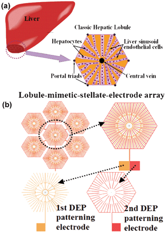
Liver Cell Patterning Lab Chip Mimicking The Morphology Of Liver Lobule Tissue Lab On A Chip Rsc Publishing from pubs.rsc.org Start studying ib biology topic 1.2: Excessive alcohol consumption can cause liver disease. The liver is both an endocrine and an exocrine gland. The hepatocyte (liver cell), the bile secretory (cholangiolar) apparatus, or the blood vascular system. Hemoglobin is degraded to iron and biliverdin, which is converted to bilirubin. The hepatic lobules are composed predominantly of liver cell trabecular cords one cell thick. June 03, 2021 reading time: The typical volume of a hepatocyte is 3.4 x 10 −9 cm 3.
The adjacent sinusoids are lined by both endothelial and kupffer cells, whereas the perisinusoidal space, located between the endothelial cells and hepatocytes, contains stellate cells and collagen fibers.
About 250 to 350 mg of unconjugated bilirubin forms daily; Ƽ store vitamins and minerals; Draw a diagram of a liver cell ib biology syllabus ib biology 231 drawing a liver cell youtube ib biology notes 23 eukaryotic cells ib biology topic 231 drawing a liver cell youtube schematic representation of copper metabolism within a liver cell. Liver cells express mscca (bear, 1990) and previous studies had shown that osmotic swelling of. The countless bile canaliculi join together into many larger bile ducts found throughout the liver. Uruj zehra mbbs, mphil, phd last reviewed: The typical volume of a hepatocyte is 3.4 x 10 −9 cm 3. The liver also stores vitamins e and k. June 03, 2021 reading time: The common hepatic duct transports the bile made by the liver cells to the gallbladder and duodenum (the first part of the small intestine) via the common bile duct. The various functions of the liver are carried out by the liver cells or hepatocytes. Insulin also activates enzymes involved in glycogen synthesis, such as glycogen synthase. Smooth endoplasmic reticulum is abundant in hepatocytes, whereas most cells in the body have only small amounts.
This allows adhesion of the second layer of cells diagram of liver. This allows adhesion of the second layer of cells.